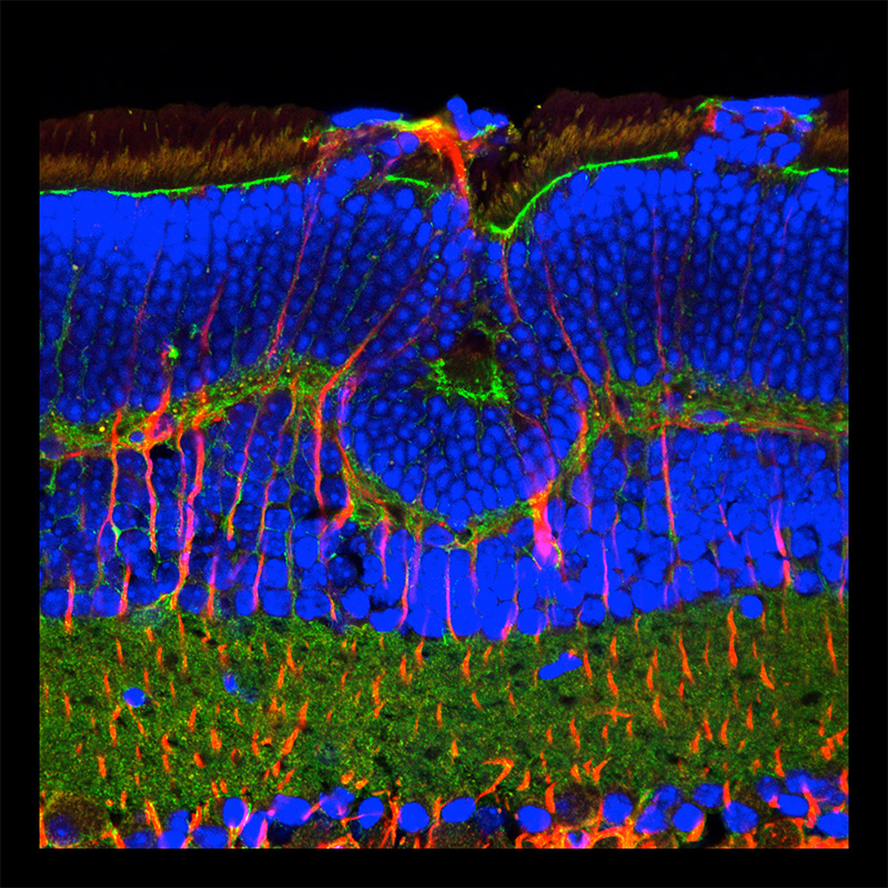
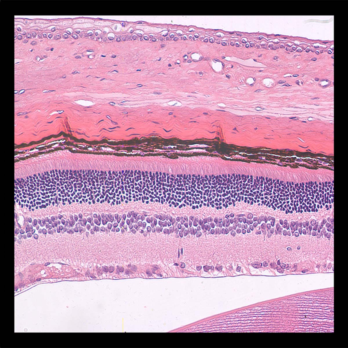
on the other side of a molecular divide (fluorescent retina)
2013
confocal micrographs | light microscopy
A series of micrographs made using confocal microscopy (stained with antibody conjugated fluorophores) and light microscopy (hematoxylin and eosin staining). These images explore the striations in rat retina where the staining allows the visualisation of different microanatomical features.

